
My research interests include:
I collaborate in these studies with George M. Martin (Dept. of Pathology)
Research Scientist, Pathology
Mailbox: 357470
Phone: 206-543-4247 in K-533 Health Sciences Bldg
Phone: 206-685-9679 in D- Health Sciences Bldg
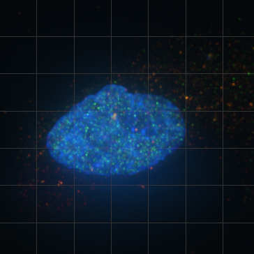
When DNA is damaged, part of the response to that damage is mobilization of gamma-H2AX. In this picture an unirradiated cell (human neuroblastoma SK-N-SH) has been fixed in 3.7% paraformaldehyde, then stained with mouse anti-gamma-H2AX (red) and rabbit anti-gamma-H2AX (green). Dapi was used to stain DNA blue.
Levels of gamma-H2AX protein are low.
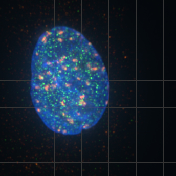
This SK-N-SH cell was irradiated with 11 Gy X-irradiation for 5.5 minutes. After 30 minutes of recovery the cell was fixed with PAF then stained for gamma-H2AX as described before.
Note that the signal from mouse anti-gamma-H2AX (red) is now prominent in large protein clusters. The signal from rabbit anti-gamma-H2AX (green) has also increased but as smaller aggregates. Why do the two signals not colocalize?
We speculate that the mouse antibody and the rabbit antibody are specific for different epitopes on the gamma-H2AX protein. When the protein is in one configuration (=I) the mouse antibody has access to its epitope while the epitope specific for the rabbit antibody is hidden. When the protein is in a different configuration (=II) the rabbit antibody has access to its epitope while that of the mouse antibody is hidden.
Assuming that the large aggregate indicate sites of DNA damage due to irradiation, configuration I may occur when gamma-H2AX is active at sites of damage, while configuration II may occur when the protein is inactive.